Chest Lung Cancer Ct Scan Images
Lung cancer detection using co learning from chest ct images and clinical demographics. Theres something called a low dose computerized tomography or low dose cat scan.
15 Images of chest lung cancer ct scan images - Are you looking for chest lung cancer ct scan images?. Make the Image Of Cat article below for as a reference or collection for your cat pictures. If you are looking for chest lung cancer ct scan images you are coming to the right page. Image Of Cat contains 15 images about chest lung cancer ct scan images, please view below.
A ct scan is usually the next test youll have after a chest x ray.
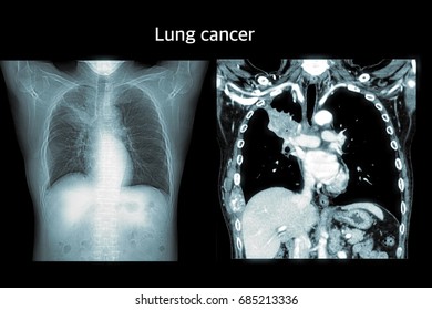
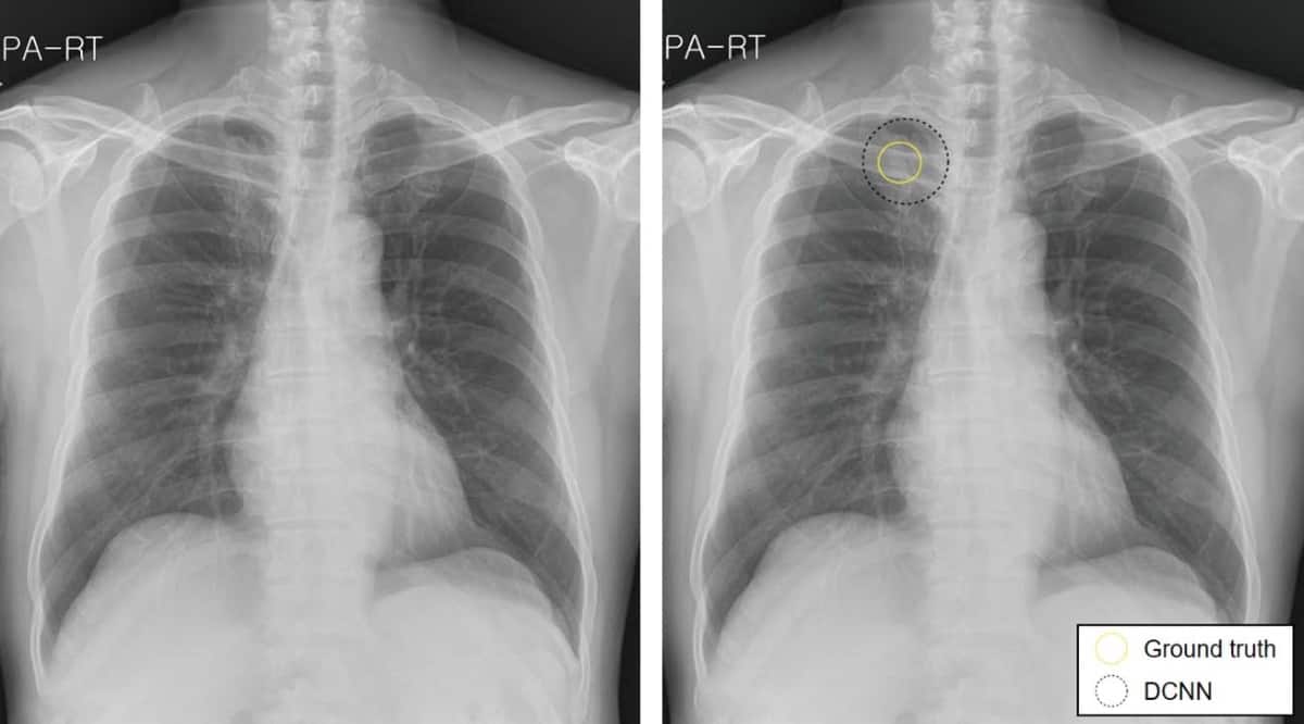
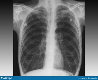
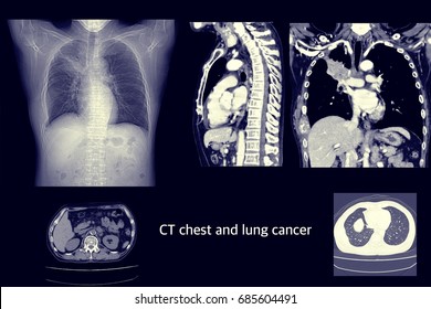
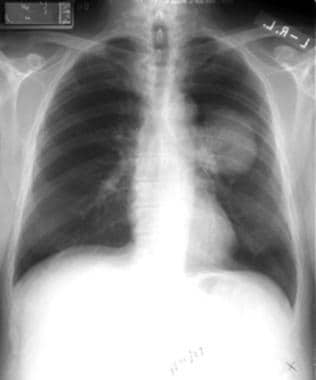
Chest lung cancer ct scan images. Computed tomography ct scan of the chest is the cornerstone of lung cancer imaging based on which further management is decided. This test can also be used to look for masses in the adrenal glands liver brain and other organs that might be due to the lung cancer spread. The mcl focuses on detection of early stage lung cancer and captures not only ct images. If a suspected area of cancer is deep within your body a ct scan might be used to guide a biopsy needle into this area to get a tissue sample to check for cancer. The bronchi are the airways that carry air to the lungs and mouth. Low dose ct scans to look for lung cancer lee health. Coloured computed tomography ct scan of a section through the chest of a 76 year old male patient with a malignant cancerous tumour bright right of the bronchus. Most lung tumours appear on x rays as a white grey mass. Computed tomography more commonly known as a ct or cat scan is a diagnostic medical imaging test. To see the difference between a blood vessel and a nodule you must scroll the pictures in the viewer frequently up and down many times. A chest x ray is usually the 1st test used to diagnose lung cancer. A computed tomography.
The cross sectional images generated during a ct scan can be reformatted in multiple planes. Many doctors used chest x rays to look for. What is ct scanning of the chest. The preprocessed whole lung scan was used as the first input channel for learning. A ct scan uses x rays and a computer to create detailed images of the inside of your body. The primary tumor shows a wide spectrum of imaging appearances. A ct scan takes a cross. Learn how lung cancer appears in x rays ct scans and. The right side of the lung is on the left side on the picture. A chest x ray is usually the first test but it cannot show that the person has cancer. Like traditional x rays it produces multiple images or pictures of the inside of the body. Though it is a very good tool in providing preliminary information about the disease it is inadequate for optimal characterization and staging.
The clear white stripes branches and spots are blood vessels. A ct scan makes mirror images. Scan is often ordered if there is something abnormal on the chest x ray. Lung cancer does not always produce symptoms in the early stages and this can make it difficult to detect.
That's 15 pictures about chest lung cancer ct scan images. Don't forget to bookmark this page for future reference, inspiration or collection cat image. Share post on Facebook / Twitter / Pinterest and others if you like this page. Thanks
