Ct Scan Images Of Brain Hemorrhage
Ct scan is almost always the first imaging modality used to assess patients with suspected intracranial hemorrhage. Left temporal epidural hematoma with a comminuted fracture of the temporal bone multiple facial fractures brain ct without contrast in brain setting bone setting.
15 Images of ct scan images of brain hemorrhage - Are you looking for ct scan images of brain hemorrhage?. Make the Image Of Cat article below for as a reference or collection for your cat pictures. If you are looking for ct scan images of brain hemorrhage you are coming to the right page. Image Of Cat contains 15 images about ct scan images of brain hemorrhage, please view below.


Tap onoff image to showhide findings.
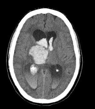
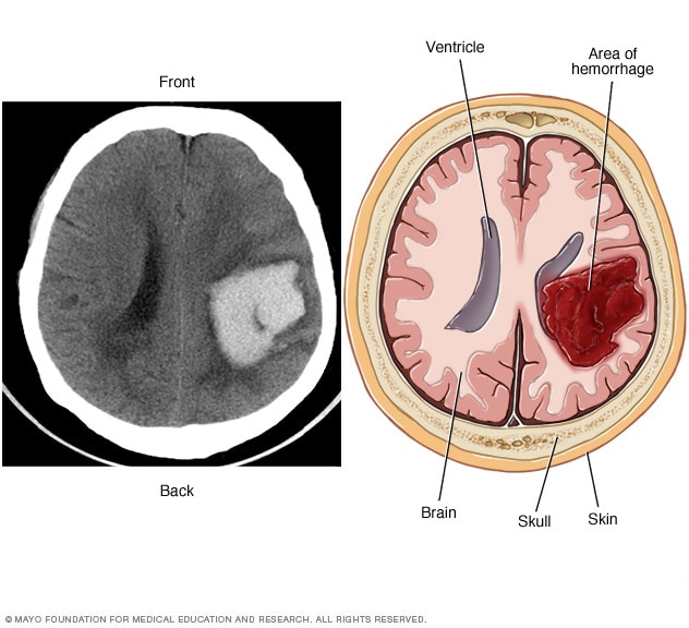
Ct scan images of brain hemorrhage. Fortunately acute blood is markedly hyperdense compared to brain parenchyma and as such usually poses little difficulty in diagnosis provided the amount of blood is large enough and the scan is performed early. Refers to a bleeding in the brain parenchyma also known as intra axial hemorrhage. When a ct scan is acquired the patient lies supine and any blood in the lateral ventricles will collect posteriorly. Ct brain images example of an intracerebral haemorrhage with extension of bleeding into the lateral ventricles. Subarachnoid haemorrhage sah blood in the ventricles may be the only sign of subarachnoid haemorrhage. Reading a ct scan in a systematic way in the emergency department can help you quickly and thoroughly assess for any neurological pathology. A ct of the brain is a noninvasive diagnostic imaging procedure that uses special x rays measurements to produce horizontal or axial images often called slices of the brain. Primary bleeding into the brain can extend into the subarachnoid space and ventricles. Max wintermark md mas the chief of neuroradiology at the university of virginia teaches you how to diagnose traumatic injuries of the brain in this dynamic video lecture. Subarachnoid haemorrhage sah hover onoff image to showhide findings. Acute hematoma is seen by pre contrast ct imaging as an area of high density. To learn more.
Ct can detect acute intracerebral blood as small as 2 mm due to contrast between high density of blood and low density of surrounding brain. Remember the mnemonic blood can be very bad follow us. There are various types of intracerebral hemorrhages see also fig. Pre contrast ct scan is the imaging procedure of choice to evaluate intracerebral hemorrhage. Brain ct scans can.


That's 15 pictures about ct scan images of brain hemorrhage. Don't forget to bookmark this page for future reference, inspiration or collection cat image. Share post on Facebook / Twitter / Pinterest and others if you like this page. Thanks

